Unveiling The Cellular Masterminds Behind Chromosome Movement: The Organelle Orchestrating Mitosis
During cell division, structures called microtubule spindles move the chromatids to opposite poles of the cell. This allows the chromatids to be separated into two new cells.
The microtubule spindle is a complex structure made up of microtubules, which are long, thin protein filaments. The microtubules are organized in a way that creates a spindle-shaped structure. The spindle fibers attach to the chromosomes at the kinetochore, which is a specialized protein complex located at the centromere of each chromosome. During cell division, the microtubule spindle shortens, pulling the chromatids to opposite poles of the cell.
The microtubule spindle is essential for cell division. Without the spindle, the chromatids would not be able to be separated into two new cells, and cell division would not be able to occur.
What Moves the Chromatids Around During Cell Division and What Organelle?
The movement of chromatids during cell division is a crucial process for ensuring the accurate distribution of genetic material to daughter cells. This process is facilitated by a specialized organelle known as the microtubule spindle. Here are six key aspects related to this topic:
- Microtubules: The building blocks of the spindle apparatus, responsible for generating the forces that move the chromosomes.
- Kinetochore: The specialized protein complex on chromosomes where spindle fibers attach, allowing for chromosome movement.
- Motor proteins: Molecules that move along microtubules, exerting pulling or pushing forces on the chromosomes.
- Centrosomes: Cellular structures that organize and nucleate microtubules, ensuring their proper bipolar orientation.
- Kinetochore checkpoints: Regulatory mechanisms that ensure proper attachment of chromosomes to the spindle before anaphase onset.
- Chromosome condensation: The process by which chromosomes become more compact and organized, facilitating their movement and segregation.
These aspects collectively contribute to the precise and efficient movement of chromatids during cell division, ensuring the faithful transmission of genetic information to future generations.
Microtubules
In the context of cell division, microtubules play a central role in the movement of chromatids. These proteinaceous structures are the fundamental components of the spindle apparatus, a complex and dynamic structure that orchestrates chromosome segregation during mitosis and meiosis.
- Structural Framework: Microtubules form the structural backbone of the spindle apparatus, providing the tracks along which chromosomes move. They are highly dynamic, constantly assembling and disassembling, allowing for the spindle to adapt to the changing needs of the cell during cell division.
- Motor Proteins: Microtubules serve as the tracks for motor proteins, such as kinesins and dyneins. These motor proteins bind to the microtubules and use the energy from ATP hydrolysis to move along them, carrying chromosomes or other cellular components.
- Force Generation: The movement of motor proteins along microtubules generates the forces that drive chromosome movement. Kinesin motors, for example, move towards the plus end of microtubules, pulling chromosomes along with them. Dynein motors, on the other hand, move towards the minus end, pushing chromosomes or other structures.
- Spindle Assembly: Microtubules are nucleated and organized by centrosomes, which are located at opposite poles of the cell. The spindle apparatus is formed by the interaction and dynamic behavior of microtubules, ensuring the proper alignment and segregation of chromosomes during cell division.
In summary, microtubules, as the building blocks of the spindle apparatus, play a critical role in generating the forces and providing the structural framework necessary for the precise movement of chromatids during cell division. Their dynamic behavior and interaction with motor proteins ensure the accurate segregation of genetic material to daughter cells.
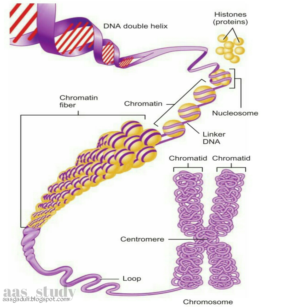
Kinetochore
The kinetochore is a crucial protein complex that serves as the attachment point between chromosomes and spindle fibers during cell division. It plays a pivotal role in ensuring the accurate segregation of chromosomes to daughter cells.
- Attachment Platform: The kinetochore provides a stable platform for the attachment of spindle fibers, which are composed of microtubules. These spindle fibers exert pulling forces on the chromosomes, enabling their movement during cell division.
- Checkpoint Control: The kinetochore is involved in cell cycle checkpoints, ensuring that all chromosomes are properly attached to spindle fibers before the cell proceeds to anaphase. This checkpoint helps prevent aneuploidy, a condition characterized by an abnormal number of chromosomes in a cell.
- Error Correction: The kinetochore can detect and correct attachment errors that may occur during spindle fiber formation. This error correction mechanism helps ensure the fidelity of chromosome segregation.
- Dynamic Structure: The kinetochore is a highly dynamic structure that undergoes significant changes during cell division. It assembles and disassembles as chromosomes attach and detach from spindle fibers, contributing to the overall regulation of chromosome movement.
In summary, the kinetochore is a specialized protein complex that plays a central role in chromosome movement during cell division. Its functions in attachment, checkpoint control, error correction, and dynamic behavior are crucial for ensuring the accurate segregation of chromosomes to daughter cells.
Motor Proteins
Motor proteins play a crucial role in the movement of chromatids during cell division. These proteins move along microtubules, which are the structural components of the spindle apparatus. The spindle apparatus is responsible for separating the chromosomes and ensuring that they are distributed equally to the daughter cells.
There are two main types of motor proteins involved in chromosome movement: kinesins and dyneins. Kinesins move towards the plus end of microtubules, while dyneins move towards the minus end. By moving along the microtubules, motor proteins exert pulling or pushing forces on the chromosomes, causing them to move towards the opposite poles of the cell.
The movement of chromosomes by motor proteins is essential for cell division. Without motor proteins, the chromosomes would not be able to separate and the cell would not be able to divide. Motor proteins are also involved in other cellular processes, such as organelle transport and cell migration.
Centrosomes
Centrosomes play a critical role in cell division by organizing and nucleating microtubules, which are essential for the movement of chromatids. Microtubules form the spindle apparatus, a structure that separates the chromosomes and ensures that they are distributed equally to the daughter cells.
- Microtubule Organization: Centrosomes organize microtubules into a bipolar spindle apparatus, with microtubules emanating from opposite poles of the cell. This bipolar orientation is crucial for the proper segregation of chromosomes during cell division.
- Microtubule Nucleation: Centrosomes nucleate microtubules, meaning they provide a site for the assembly of new microtubules. This nucleation process is essential for the formation of the spindle apparatus and the subsequent movement of chromosomes.
- Chromosome Attachment: Centrosomes serve as the primary site for the attachment of spindle fibers to chromosomes. This attachment is mediated by specialized protein complexes called kinetochores, which ensure that chromosomes are properly aligned and segregated during cell division.
- Cell Cycle Regulation: Centrosomes are also involved in regulating the cell cycle, particularly the transition from G2 phase to mitosis. They ensure that all chromosomes are properly attached to the spindle apparatus before anaphase onset, preventing errors in chromosome segregation.
In summary, centrosomes play a critical role in cell division by organizing and nucleating microtubules, ensuring their proper bipolar orientation. These functions are essential for the accurate segregation of chromosomes to daughter cells, highlighting the importance of centrosomes in maintaining genomic stability and cell viability.
Kinetochore checkpoints
Kinetochore checkpoints are crucial regulatory mechanisms that ensure the accurate segregation of chromosomes during cell division. Their role is tightly intertwined with the cellular processes involved in "what moves the chromatids around during cell division and what organelle".
- Monitoring Attachment: Kinetochore checkpoints monitor the proper attachment of chromosomes to the spindle apparatus, which is essential for faithful chromosome segregation. This monitoring ensures that all chromosomes are correctly aligned and attached before the cell proceeds to anaphase, the stage where sister chromatids are separated and pulled to opposite poles of the dividing cell.
- Preventing Premature Separation: By halting the cell cycle progression until all chromosomes are properly attached, kinetochore checkpoints prevent premature separation of sister chromatids. This prevention minimizes the risk of aneuploidy, a condition characterized by an abnormal number of chromosomes in a cell, which can lead to developmental abnormalities or diseases.
- Error Correction: Kinetochore checkpoints also play a role in error correction. If a chromosome is not properly attached to the spindle, the checkpoint can trigger mechanisms to correct the attachment or, in some cases, eliminate the improperly attached chromosome to prevent its missegregation.
- Coordination with Motor Proteins: Kinetochore checkpoints coordinate with motor proteins, such as kinesins and dyneins, which are responsible for moving chromosomes along the spindle fibers. This coordination ensures that chromosomes are moved to the correct poles of the dividing cell, further contributing to accurate chromosome segregation.
In summary, kinetochore checkpoints are essential regulatory mechanisms that work in conjunction with the cellular machinery involved in chromosome movement during cell division. These checkpoints ensure that chromosomes are properly attached to the spindle apparatus, preventing premature separation and promoting accurate chromosome segregation. Their role is critical for maintaining genomic stability and preventing aneuploidy, highlighting their importance in the context of "what moves the chromatids around during cell division and what organelle".
Chromosome condensation
Chromosome condensation is a crucial process that occurs during cell division and plays a pivotal role in facilitating the movement and segregation of chromosomes. It involves the compaction and organization of the chromatin fibers that make up chromosomes, resulting in structures that are more condensed and easier to separate during cell division.
The condensation of chromosomes is essential for the accurate segregation of genetic material to daughter cells. Highly condensed chromosomes are less prone to entanglement and breakage, ensuring that each daughter cell receives a complete and intact set of chromosomes. The process of chromosome condensation is mediated by a protein complex called condensin, which binds to the chromatin fibers and introduces loops and coils, leading to the compaction of the chromosome.
The timing of chromosome condensation is precisely regulated and coordinated with other events during cell division. Condensation typically begins in prophase, the early stage of mitosis, and reaches its maximum level in metaphase, when the chromosomes are aligned at the metaphase plate. This condensation facilitates the interaction of chromosomes with the spindle fibers, which are responsible for separating the chromosomes and moving them to opposite poles of the cell during anaphase.
The process of chromosome condensation is essential for the accurate and successful completion of cell division. It ensures the proper segregation of genetic material, which is critical for maintaining the genetic integrity of the cell and preventing chromosomal abnormalities. Understanding the mechanisms and regulation of chromosome condensation is therefore of great importance in the field of cell biology and has implications for our understanding of cell division, genetic disorders, and developmental biology.
FAQs on "What Moves the Chromatids Around During Cell Division and What Organelle?"
This section addresses frequently asked questions related to the topic of chromosome movement during cell division, providing concise and informative answers to enhance understanding.
Question 1: What is the primary structure responsible for moving chromosomes during cell division?
Answer: Microtubules, organized into a structure called the spindle apparatus, are primarily responsible for moving chromosomes during cell division.
Question 2: What is the role of motor proteins in chromosome movement?
Answer: Motor proteins, such as kinesins and dyneins, move along microtubules and exert forces on chromosomes, facilitating their movement towards opposite poles of the cell.
Question 3: How does the kinetochore contribute to chromosome movement?
Answer: The kinetochore serves as the attachment point between chromosomes and spindle fibers, ensuring proper chromosome alignment and segregation during cell division.
Question 4: What is the significance of centrosomes in chromosome movement?
Answer: Centrosomes play a crucial role in organizing and nucleating microtubules, establishing the bipolar spindle apparatus essential for chromosome movement and segregation.
Question 5: How do kinetochore checkpoints ensure accurate chromosome segregation?
Answer: Kinetochore checkpoints monitor proper chromosome attachment to the spindle apparatus, preventing premature separation and promoting faithful chromosome segregation.
Question 6: What is the purpose of chromosome condensation during cell division?
Answer: Chromosome condensation compacts and organizes chromosomes, making them more manageable for movement and segregation during cell division.
In summary, the coordinated action of microtubules, motor proteins, kinetochores, centrosomes, kinetochore checkpoints, and chromosome condensation ensures the precise and efficient movement of chromosomes during cell division, a fundamental process for maintaining genetic integrity and cellular function.
Transition to the next article section: Next Section
Tips on Understanding "What Moves the Chromatids Around During Cell Division and What Organelle"
Grasping the intricacies of chromosome movement during cell division requires a comprehensive approach. Here are some valuable tips to enhance your understanding:
Tip 1: Visualize the Process: Create mental images or diagrams depicting the spindle apparatus, microtubules, motor proteins, and chromosomes. This visual representation aids in comprehending the dynamic interactions involved.
Tip 2: Focus on the Kinetochore's Role: Recognize the kinetochore's critical function as the attachment point between chromosomes and spindle fibers. Understanding its structure and mechanisms helps unravel the precise chromosome segregation process.
Tip 3: Explore Motor Protein Diversity: Familiarize yourself with the different types of motor proteins, such as kinesins and dyneins. Comprehend their roles in exerting pulling or pushing forces that facilitate chromosome movement.
Tip 4: Appreciate Centrosome Function: Understand the significance of centrosomes in organizing microtubules and establishing the bipolar spindle apparatus. This knowledge illuminates how centrosomes contribute to the overall chromosome movement process.
Tip 5: Study Kinetochore Checkpoint Mechanisms: Delve into the mechanisms of kinetochore checkpoints. Learn how they ensure proper chromosome attachment, preventing errors that could lead to aneuploidy.
Tip 6: Examine Chromosome Condensation: Comprehend the process of chromosome condensation and its impact on chromosome structure. Recognize its importance in facilitating chromosome movement and segregation.
Tip 7: Utilize Resources and Literature: Consult textbooks, research papers, and reputable online resources to supplement your understanding. Engage with experts in the field to gain deeper insights and clarify any uncertainties.
Key Takeaways: By implementing these tips, you will enhance your grasp of the mechanisms underlying chromosome movement during cell division. This knowledge is essential for comprehending the fundamental processes that ensure accurate genetic inheritance.
Transition to Conclusion: As you delve deeper into the topic, remember that a comprehensive understanding of chromosome movement not only illuminates the intricacies of cell division but also provides a foundation for exploring broader concepts in genetics and cell biology.
Conclusion
This in-depth exploration of "what moves the chromatids around during cell division and what organelle" has shed light on the intricate mechanisms that ensure accurate chromosome segregation during cell division. We have examined the critical roles of microtubules, motor proteins, kinetochores, centrosomes, kinetochore checkpoints, and chromosome condensation in orchestrating this fundamental process.
Understanding the principles governing chromosome movement not only provides a deeper appreciation for the complexity of cell division but also lays the groundwork for future research endeavors in genetics and cell biology. By unraveling the molecular mechanisms behind chromosome segregation, scientists can gain valuable insights into the causes and potential treatments for genetic disorders and diseases.
Uncover The Mystery: When Did Steve Young Tie The Knot?
Brent Faiyaz's Race: Exploring Identity, Fluidity, And Impact
Is Ross Chastain Married? Find Out Here!


